Ewingella americana
What does the cancer research actually show?
Editorial orientation / 3 minute read
Ewingella americana: reading the mouse study
Ewingella americana is a bacterium, not a frog secretion or an approved cancer treatment. The study behind this dossier, by Seigo Iwata and colleagues in Gut Microbes (2025), investigated bacteria isolated from amphibian and reptile intestines. Its striking result was obtained in a mouse tumour model. Bench now distinguishes that experiment from the much larger claim that a bacterium could treat human cancer.
© 2025 Iwata et al., Gut Microbes 17(1), 2599562. CC BY-NC 4.0. Figure unaltered.
What was actually studied
The investigators screened bacterial isolates, then examined selected strains in cell experiments and mice. The principal animal model used Colon26 mouse colorectal-cancer cells in an immunocompetent mouse system. The tumours were implanted under the skin; this is not the same biological setting as a tumour developing inside a human colon. The work included three-dimensional cancer-cell spheroids, tumour-growth measurements, tissue imaging, immune measurements and safety observations.
In the illustrated comparison, the E. americana group had five animals and the authors reported complete tumour responses. The comparison included anti-PD-L1 treatment, liposomal doxorubicin and a saline control. That finding concerns the study's specific model and comparators. It does not establish superiority over the full range of clinical colorectal-cancer treatments.
The paper also reports a separate tumour rechallenge experiment, intended to investigate whether treated animals retained protective immune memory. Such experiments address a different question from initial tumour shrinkage and should not be folded into one undifferentiated “cure rate.”
The proposed two-part explanation
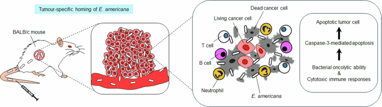
The authors propose both direct damage to cancer cells and recruitment of the host immune response. Their interpretation includes bacterial accumulation in tumours, local proliferation, tumour-cell death and increased involvement of immune cells. The paper examines T cells, B cells and neutrophils alongside inflammatory signals.
A mechanism drawing is a useful map of this interpretation. It is not itself an additional experiment. To assess the mechanism, distinguish the observed measurements—such as bacterial counts or stained tissue—from the causal story that connects them. Several biological processes can occur together without every arrow in a proposed pathway having been independently established.
How to read the figures
Tumour-growth curves describe changes over the observation period. The sample size belongs with the curve: a complete response in five animals is a small preclinical result, not an estimate of a human treatment-success rate. Error bars describe variation under the reported analysis; they do not capture all uncertainty about translation to another model or species.
Tissue images answer a different kind of question. They can illustrate where signals or cells were detected, but interpretation depends on the marker, control and sampling. Compare them with the authors' quantitative analyses instead of treating a visually dramatic image as a clinical outcome.
The locally preserved illustrations are attributed to the original research. Their scientific labels have been retained, and Bench's captions identify the animal-study context. The source room contains a selected results-and-discussion record so the guide can be checked without leaving Bench.
Safety and translation remain research questions
The paper reports clearance and toxicity observations in mice. Those observations have boundaries: animal strain, tumour model, measurement method, observation interval and sample size. They do not establish that administering the organism to people is safe. A strain used in a laboratory experiment is not interchangeable with an organism recovered outside that controlled setting.
The authors discuss additional tumour models and clinical-development work as future research. Questions include reproducibility, containment of infection, immune responses, distribution outside tumours, interactions with other treatments and manufacturing consistency. This dossier describes the research rather than supplying an administration protocol.
What the result does—and does not—support
The study supports further investigation of a particular bacterial strain as a preclinical anticancer candidate. It supplies substantially more than a headline or an in-vitro-only observation. It does not supply human trial results or establish a general cure for cancer. Those two distinctions should remain visible together.
The original selected archive address was unavailable. This reading guide therefore uses the primary paper, DOI 10.1080/19490976.2025.2599562, and JAIST's accompanying research announcement. It is a Bench editorial guide; the article's authors remain the source of the experimental claims.
Source notes & attribution
- https://www.jaist.ac.jp/english/whatsnew/press/2025/12/17-1.html
- https://pmc.ncbi.nlm.nih.gov/articles/PMC12710904/
Go deeper.
Primary reading25 min
Ewingella americana — primary results and discussion
© 2025 Iwata et al. Gut Microbes 17(1), 2599562. CC BY-NC 4.0; selected Results and Discussion text, with laboratory protocols omitted. Figures retain the authors’ claims and labels.
The visual record.
3 archived figures · open to inspectThe source room.
Everything readable here stays on StrangewellArchived texts preserve what the source said. They may contain historical, speculative, or promotional claims; inclusion is not verification.
Source text25 min
Ewingella americana — primary results and discussion
© 2025 Iwata et al. Gut Microbes 17(1), 2599562. CC BY-NC 4.0; selected Results and Discussion text, with laboratory protocols omitted. Figures retain the authors’ claims and labels.